

Craneofacial manifestaciones in patients with renal osteodystrophy
Keywords:
chronic renal failure; mineral and bone disorder; chronic kidney disease; secondary hyperparathyroidism; hyperphosphatemia; hypercalcemia.Abstract
Introduction: Renal osteodystrophy is a diffuse metabolic osteopathy, related to chronic renal failure, which includes various pathologies in the musculoskeletal system. It occurs in response to metabolic disorders generated by electrolyte changes, chronic inflammation and hormonal alteration. These variations modify the process of bone remodeling. Clinical manifestations include alteration in the parenchyma and bone stroma, and range from expansive lesions, producing deformity, to pathological fractures of the bone.
Objective: Relate the clinical, imaging and histological manifestations in the skull-facial bone component for the diagnosis of renal osteodystrophy.
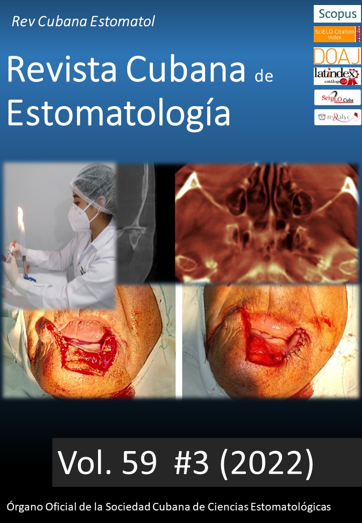
Case Presentation: Two clinical cases of patients with chronic kidney disease and secondary underlying hyperparathyroidism are presented, with multiple masses in the jaws that cause facial asymmetry and functional alteration. Tomographic images reveal alteration in cortical and trabecular bone morphology. Both individuals showed alterations in levels of parathyroid hormone, alkaline phosphatase, phosphorus and serum calcium. Histopathology verified fibro-bone tissue with neoformed bone and great vascularization, with multinucleated osteoclast-like cells without the presence of hemosiderin. Patients were attended in an interdisciplinary way between internal medicine, endocrinology and maxillofacial surgery.
Conclusions: Patients with advanced chronic kidney disease present alteration of the structure and bone and mineral metabolism. Such a situation can compromise the craniofacial bone complex. Severe cases of renal osteodystrophy are characterized by a marked expansion of the jaws, which generates asymmetry and traits of leonthiasis. The tomographic images associated with renal osteodystrophy present trabecular bones with cortical expansion, which evidences the immature bone turnover present. Histopathology is not specific and may be similar to cases of craniofacial bone dysplasia. Given the above, it is essential to relate these findings to the clinic to define an adequate diagnosis.Downloads
References
Cannata J, Gómez C, Grosso S, et al. Osteodistrofia renal: clasificación y concepto actual. Nefrología. 1995;15(1):20-24. https://www.revistanefrologia.com/es-osteodistrofia-renal-clasificacion-concepto-actual--articulo-X0211699595022756
Astudillo J, Cocio R, Ríos D. Osteodistrofia renal y trastornos del metabolismo y la mineralización ósea asociados a enfermedad renal crónica: manifestaciones en radiología. Rev ChilRad. 2016; 22(1): 27-34. https://www.sciencedirect.com/science/article/pii/S0717201X16000075
Moe S, Drüeke T, Cunningham J, Goodman W, et al. Definition, evaluation, and classification of renal osteodystrophy: A position statement from Kidney Disease: Improving Global Outcomes (KDIGO). Kidney Int. 2006 Jun; 69(11):1945-53. https://www.kidney-international.org/article/S0085-2538(15)51415-4/fulltext
Lorenzo V, Rodríguez M, Pérez R y Cannata J. De la osteodistrofia renal a las alteraciones Del metabolismo óseo y mineral asociado a la enfermedad renal crónica: evolución de un concepto. Nefrología. 2007; 27(5):527-533. https://www.revistanefrologia.com/es-de-osteodistrofia-renal-alteraciones-del-articulo-X0211699507021867
Bover J, et al. Osteoporosis, densidad mineral ósea y complejo CKD-MBD (I): consideraciones diagnósticas. Nefrología Sept – Oct 2018; 38 (5): 476 – 490. https://www.revistanefrologia.com/es-osteoporosis-densidad-mineral-osea-complejo-ckd-mbd-i-consideraciones-diagnosticas-articulo-S0211699518300444
Bakkaloglu S, Wesseling-Perry K, Pereira RC, Gales B, Wang H, Elashoff R, Salusky I. Value of the new bone classification system in pediatric renal osteodystrophy. Clin J Am Soc Nephrol. 2010; 5(10):1860-6. https://cjasn.asnjournals.org/content/5/10/1860.long
De Sousa M, et al. Severe maxillofacial renal osteodystrophy in two patients with chronic kidney disease. Oral Maxillofac Surg (2015) 19:321–327. https://link.springer.com/article/10.1007%2Fs10006-015-0490-9
Kaushik A, Kaushik M. Maxillofacial radiographic changes in renal osteodystrophy. Journal of Parathyroid Disease March 2016, 4(1), 13–16. http://jparathyroid.com/Article/JPD_20160410104447
Raubenheimer EJ, Noffke CE, Mohamed A. Expansive jaw lesions in chronic kidney disease: review of the literature and a report of two cases. Oral Surg Oral Med Oral Pathol Oral Radiol. 2015 Mar; 119(3):340-5. https://www.oooojournal.net/article/S2212-4403(14)01360-1/fulltext
Corrêa FS y cols. Oral and maxillofacial manifestations of chronic kidney disease–mineral and bone disorder: a multicenter retrospective study. Oral Surg Oral Med Oral Pathol Oral Radiol. 2018 Jan; 125(1):31-43. https://www.oooojournal.net/article/S2212-4403(17)31067-2/fulltext
Davis EM. Oral Manifestations of Chronic Kidney Disease and Renal Secondary Hyperparathyroidism: A Comparative Review. J Vet Dent. Summer 2015; 32(2):87-98. https://journals.sagepub.com/doi/abs/10.1177/089875641503200202
James BC, Hwang JL, Grogan RH, Kaplan EL, Sarne D, Angelos P. Leontiasis ossea caused by long-standing hyperparathyroidism secondary to chronic renal failure. Surgery. 2014;156(6): 1644-6. https://www.surgjournal.com/article/S0039-6060(14)00560-1/fulltext
Haroyan H, Bos A, Ginat DT. Uremic leontiasis ossea. Am J Otolaryngol. 2015;36(1):74-6. https://www.sciencedirect.com/science/article/abs/pii/S0196070914001914
Donoso-Hofer F, Gunther-Wood M, Romero-Romano P, Pezoa-Opazo N, Fernández-Toro MA, Ortega-Pinto AV. Uremic leontiasis ossea, a rare presentation of severe renal osteodystrophy secondary to hyperparathyroidism. J Stomatol Oral Maxillofac Surg. 2018;119(1):56-60. https://www.sciencedirect.com/science/article/abs/pii/S2468785517301842?via%3Dihub
Moorthi RN, Moe SM. Recent advances in the noninvasive diagnosis of renal osteodystrophy. Kidney Int. 2013;84(5):886-94. https://www.kidney-international.org/article/S0085-2538(15)56074-2/fulltext
Published
How to Cite
Issue
Section
License
Authors retain all rights to their works, which they can reproduce and distribute as long as they cite the primary source of publication.
The Rev Cubana Estomatol is subject to the Creative Commons Attribution-Non-Commercial 4.0 International License (CC BY-NC 4.0) and follows the publication model of SciELO Publishing Schema (SciELO PS) for publication in XML format.
You are free to:
- Share — copy and redistribute the material in any medium or format.
- Adapt — remix, transform, and build upon the material.
The licensor cannot revoke these freedoms as long as you follow the license terms.
Under the following terms:
Attribution — You must give appropriate credit, provide a link to the license, and indicate if changes were made. You may do so in any reasonable manner, but not in any way that suggests the licensor endorses you or your use.
- NonCommercial — You may not use the material for commercial purposes.
No additional restrictions — You may not apply legal terms or technological measures that legally restrict others from doing anything the license permits.
Notices:
- You do not have to comply with the license for elements of the material in the public domain or where your use is permitted by an applicable exception or limitation.
- No warranties are given. The license may not give you all of the permissions necessary for your intended use. For example, other rights such as publicity, privacy, or moral rights may limit how you use the material.