

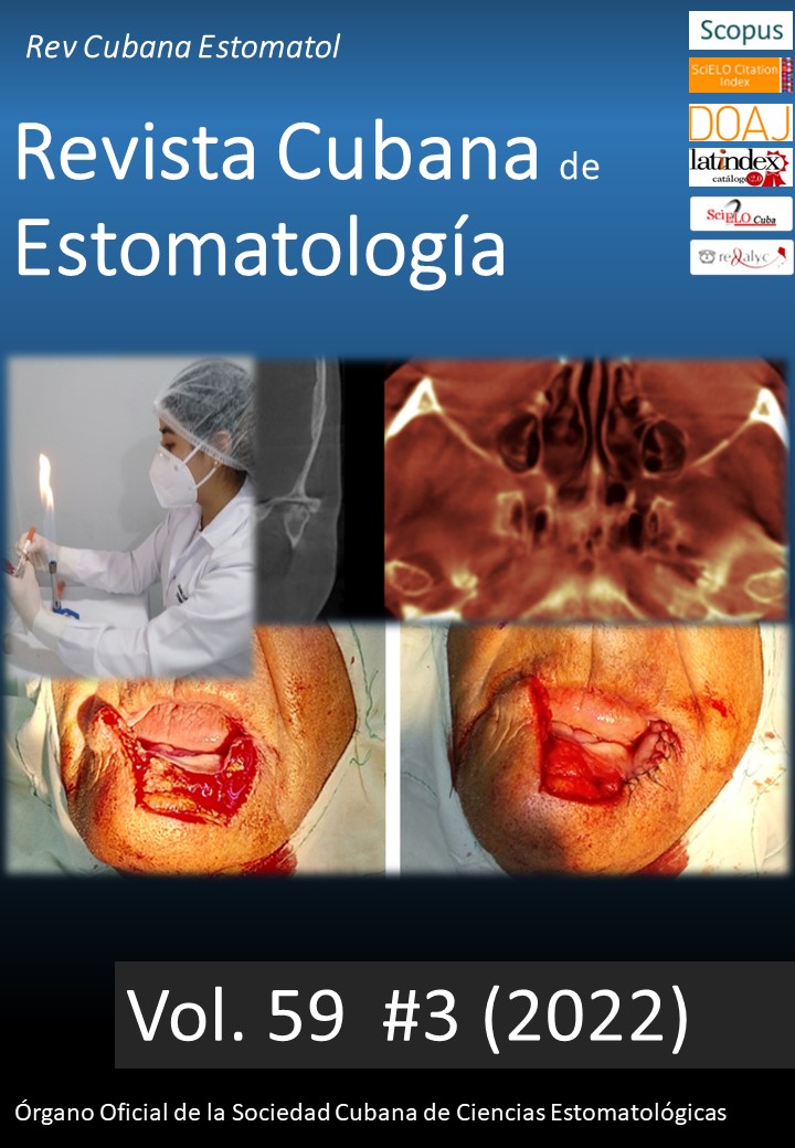
Fossa navicularis magna: detection by cone-beam computed tomography
Keywords:
Cone-beam computed tomography; skull base; fossa navicularis magna; medium basilar channel.Abstract
Introduction: The identification, interpretation and management of incidental findings in dental imaging is essential. Some of them require additional imaging techniques and referral to experienced professionals, others only their description. One of these anatomical variants is found in the clivus, fossa navicularis magna, associated in few cases with systemic repercussions.
Objective: Describe the characteristics of fossa navicularismagna for its identification by cone-beam computed tomography.
Presentation of cases: Three female patients, with an age range between 35-71 years who come to the Dental Center of San Martín de Porres University for orthodontic treatments and oral rehabilitation. In these areas, as part of the protocol, cone-beam computed tomography per retained piece and development of surgical guides are requested, respectively. The scan allows the identification of a notch-like defect in the clivus, of well-defined boundaries and corticalized edges, suggesting fossa navicularis magna. The patients' medical history did not suggest clinical implications.
Main comments: This anatomical variant is reported and discussed and its presence does not require treatment and generally has no systemic repercussions. In rare cases it has been associated with clinical pictures that threaten the patient's life, precisely because it can serve as a path for intracranial infections. Hence the need to know and describe this anatomical variant.
Downloads
References
Neelakantan A, Rana AK. Benign and malignant diseases of clivus. Clin Radiol; 2014 [acceso 22/12/2019];69(12):1295-303. Disponible en: https://pubmed.ncbi.nlm.nih.gov/25168701/
Ray B, Kalthur SG, Kumar B, Bhat MR, D’souza AS, Gulati HS, et al. Morphological variations in the basioccipital region of the South Indian skull. Nepal J Med Sci; 2014 [acceso 24/12/2019];3(2):124-8. Disponible en: (PDF) Morphological variations in the basioccipital region of the South Indian skull (researchgate.net)
Segal N, Atamne E, Shelef I, Zamir S, Landau D. Intracranial infection caused by spreading through the fossa navicularis magna - A case report and review of the literature. Int J Pediatr Otorhinolaryngol; 2013 [acceso 26/05/2020];77(12):1919-21. Disponible en: https://pubmed.ncbi.nlm.nih.gov/24148862/
Syed AZ, Mupparapu M. Fossa navicularis magna detection on cone-beam computed tomography. Imaging Sci Dent; 2016 [acceso 26/05/2020];46(1):47-51. Disponible en: https://pubmed.ncbi.nlm.nih.gov/27051639/
Cankal F, Ugur HC, Tekdemir I, Elhan A, Karahan T, Sevim A. Fossa navicularis: anatomic variation at the skull base. Clin Anat; 2004 [acceso 25/05/2020];17(2):118-22. Disponible en: https://pubmed.ncbi.nlm.nih.gov/14974099/
Prabhu SP, Zinkus T, Cheng AG. Clival osteomyelitis resulting from spread of infection through the fossa navicularis magna in a child. Pediatr Radiol; 2009 [acceso 12/01/2020]; 39:995-8. Disponible en: https://pubmed.ncbi.nlm.nih.gov/19415254/
Alsufyani NA. Cone beam computed tomography incidental findings of the cervical spine and clivus: retrospective analysis and review of the literature. Oral Surg Oral Med Oral Pathol Oral Radiol; 2017 [acceso 26/05/2020] Jun;123(6):e197-e217. Disponible en: https://www.oooojournal.net/article/S2212-4403(17)30089-5/fulltext
Ersan N. Prevalence and morphometric features of fossa navicularis on cone beam computed tomography in Turkish population. Folia Morphol; 2017 [acceso 20/01/2020];76(4):715-9. Disponible en: https://pubmed.ncbi.nlm.nih.gov/28353302/
Ginat DT, Ellika SK, Corrigan J. Multi-Detector-Row Computed Tomography Imaging of Variant Skull Base Foramina. J Comput Assist Tomogr; 2013 [acceso 22/01/2020]; 37(4):481-5. Disponible en: https://pubmed.ncbi.nlm.nih.gov/23863520/
De Vos W, Casselman J, Swennen GRJ. Cone-beam computerized tomography (CBCT) imaging of the oral and maxillofacial region: A systematic review of the literature. Int. J Oral Maxillofac Surg; 2009 [acceso 22/01/2020];38: 609–25. Disponible en: https://www.ijoms.com/article/S0901-5027(09)00864-9/fulltext
Sheikh S, Iwanaga J, Rostad S, Rustagi T, Oskouian RJ, Tubbs RS. The First Histological Analysis of the Tissues Lining the Fossa Navicularis: Insights to its Etiology Cureus; 2017 [acceso 25/05/2020];9(5):e1299. Disponible en: https://europepmc.org/article/pmc/pmc5493477
Conley LM, Phillips CD. Imaging of the Central Skull Base. Radiol Clin North Am: 2017 [acceso 26/05/2020];55(1):53-67. Disponible en: https://pubmed.ncbi.nlm.nih.gov/27890188/
Ersan AP. Prevalence of fossa navicularis among cleft palate patients detected by cone beam computed tomography. Yeditepe Dental Journal; 2017 [acceso 26/05/2020];13:21-3. Disponible en: https://www.researchgate.net/publication/316230672_Prevalence_of_fossa_navicularis_among_cleft_palate_patients_detected_by_cone_beam_computed_tomography
Bayrak S, Göller Bulut D, Orhan K. Prevalence of anatomical variants in the clivus: fossa navicularis magna, canalis basilaris medianus, and craniopharyngeal canal. Surg Radiol Anat; 2019 [acceso 10/01/2020];41(4):477-83. Disponible en: https://pubmed.ncbi.nlm.nih.gov/30725217/
Kaplan FA, Yesilova E, Bayrakdar IS, Ugurlu M. Evaluation of the relationship between age and gender of fossa navicularis magna with cone-beam computed tomography in orthodontic subpopulation. J Anat Soc India; 2019 [acceso 05/01/2020];68:201-4. Disponible en: http://www.jasi.org.in/article.asp?issn=0003-2778;year=2019;volume=68;issue=3;spage=201;epage=204;aulast=Kaplan
Magat G. Evaluation of morphometric features of fossa navicularis using cone-beam computed tomography in a Turkish subpopulation. Imaging Sci Dent; 2019 [acceso 05/01/2020] Sep;49(3):209-12. Disponible en: https://www.ncbi.nlm.nih.gov/pmc/articles/PMC6761062/
Syed AZ, Zahedpasha S, Rathore SA, Mupparapu M. Evaluation of canalis basilaris medianus using cone-beam computed tomography. Imaging Sci Dent; 2016 [acceso 26/05/2020];46(2):141-4. Disponible en: https://www.ncbi.nlm.nih.gov/pmc/articles/PMC4925651/
Chandra T, Maheshwari M, Kelly TG, Segall HD, Agarwal M, Mohan S. Imaging of Pediatric Skull Base Lesions. Neurographics; 2015 [acceso 26/05/2020];5(2):72–84. Disponible en: https://www.researchgate.net/publication/273525949_Imaging_of_Pediatric_Skull_Base_Lesions
Miyahara H, Matsunaga T. Tornwaldt’s disease. Acta Otolaryngol Suppl; 1994 [acceso 25/05/2020]; 517:36-9. Disponible en: https://www.tandfonline.com/doi/abs/10.3109/00016489409124336?journalCode=ioto20
Chong VFH, Fan YF. Radiology of the nasopharynx: pictorial essay. Australas Radiol; 2000 [acceso 26/05/20202];44:5–13. Disponible en: https://onlinelibrary.wiley.com/doi/abs/10.1046/j.1440-1673.2000.00765.x?sid=nlm%3Apubmed
Beltramello A, Puppini G, El-Dalati G, Girelli M, Cerini R, Sbarbati A, Pacini P. Fossa navicularis magna. Am J Neuroradiol; 1998 [acceso 26/05/2020]; 19(9):1796–8. Disponible en: http://www.ajnr.org/content/19/9/1796.long
Lohman BD, Sarikaya B, McKinney AM, Hadi M. Not the typical Tornwaldt’s cyst this time? A nasopharyngeal cyst associated with canalis basilaris medianus. Br J Radiol; 2011 [acceso 26/05/2020];84:e169-71. Disponible en: https://www.birpublications.org/doi/full/10.1259/bjr/95083086?url_ver=Z39.88-2003&rfr_id=ori:rid:crossref.org&rfr_dat=cr_pub%20%200pubmed
Benadjaoud Y, Klopp-Dutote N, Choquet M, Brunel E, Guiheneuf R, Page C. A case of acute clival osteomyelitis in a 7-year-old boy secondary to infection of a Thornwaldt cyst. Int J Pediatr Otorhinolaryngol; 2017 [acceso 26/05/2020];95:87-90. Disponible en: https://europepmc.org/article/med/28576541
Alalade AF, Briganti G, Mckenzie JL, Gandhi M, Amato D, Panizza BJ, et al. Fossa navicularis in a pediatric patient: anatomical skull base variant with clinical implications. J Neurosurg Pediatr; 2018 [acceso 26/05/2020];22(5):523-7. Disponible en: https://thejns.org/pediatrics/view/journals/j-neurosurg-pediatr/22/5/article-p523.xml
Kunimatsu A, Kunimatsu N. Skull Base Tumors and Tumor-Like Lesions: A Pictorial Review. Pol J Radiol; 2017 [acceso 26/05/2020];82:398–409. Disponible en: https://pubmed.ncbi.nlm.nih.gov/28811848/
Published
How to Cite
Issue
Section
License
Authors retain all rights to their works, which they can reproduce and distribute as long as they cite the primary source of publication.
The Rev Cubana Estomatol is subject to the Creative Commons Attribution-Non-Commercial 4.0 International License (CC BY-NC 4.0) and follows the publication model of SciELO Publishing Schema (SciELO PS) for publication in XML format.
You are free to:
- Share — copy and redistribute the material in any medium or format.
- Adapt — remix, transform, and build upon the material.
The licensor cannot revoke these freedoms as long as you follow the license terms.
Under the following terms:
Attribution — You must give appropriate credit, provide a link to the license, and indicate if changes were made. You may do so in any reasonable manner, but not in any way that suggests the licensor endorses you or your use.
- NonCommercial — You may not use the material for commercial purposes.
No additional restrictions — You may not apply legal terms or technological measures that legally restrict others from doing anything the license permits.
Notices:
- You do not have to comply with the license for elements of the material in the public domain or where your use is permitted by an applicable exception or limitation.
- No warranties are given. The license may not give you all of the permissions necessary for your intended use. For example, other rights such as publicity, privacy, or moral rights may limit how you use the material.