

Enucleation of tooth cyst, associated with retained lateral incisor, and orthodontic traction of retained upper canine
Keywords:
tooth cyst, impacted tooth, enucleation.Abstract
Introduction: A drawback of permanent tooth eruption is tooth retention. A retained tooth has not completed its eruption and has not reached its normal position in the jaw. Retained teeth may be within the jaw asymptomatic or causing tooth migration, persistence of deciduous teeth, alterations in occlusion and aesthetics, formation of tooth cyst and tumors.
Objective: Describe the enucleation of dentiger cyst, associated with the lateral incisor, and orthodontic traction of retained upper canine.
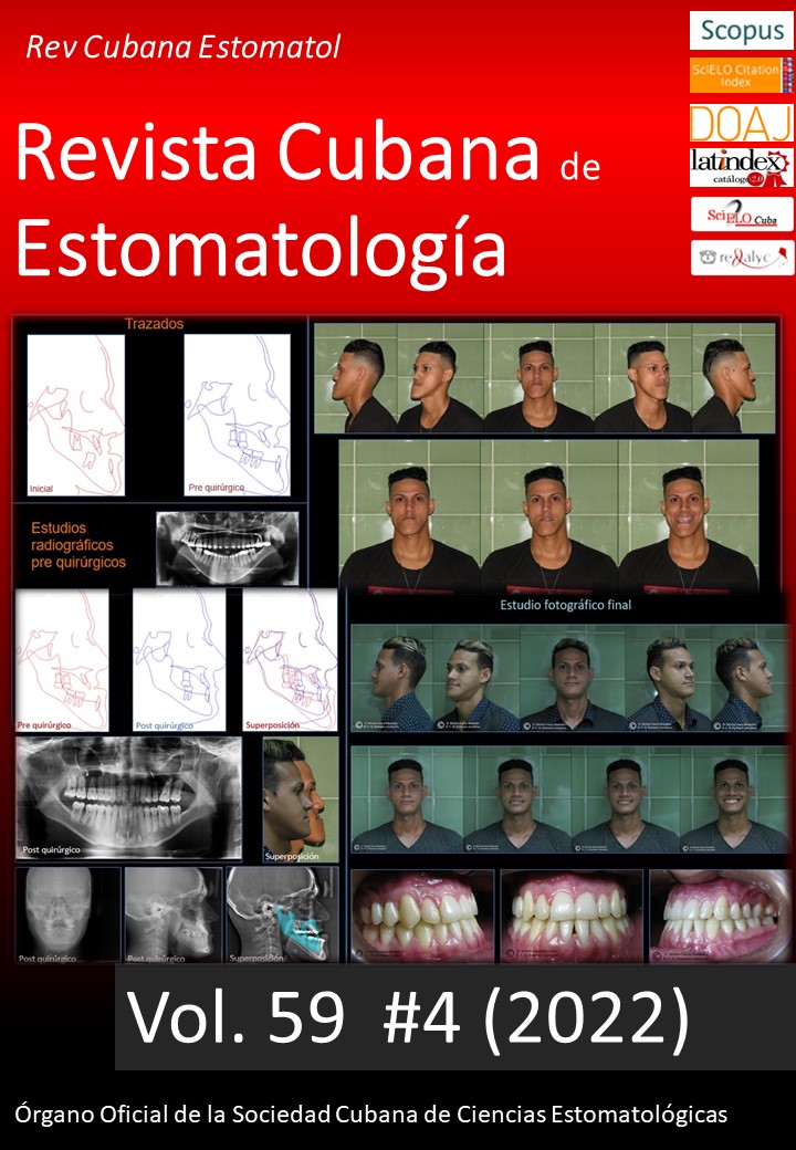
Case presentation: Female patient, 19 years old, with persistence of teeth 52 and 53. Tomographically there is a right upper lateral incisor retained in a horizontal position, associated with a radiolucent image of dimensions of 11.2 mm x 20.1 mm and a canine retained in an upright position. Exodontics were performed at the flap of the lateral incisor and enucleation of the cyst. Regarding the canine, the orthodontic button was placed for subsequent traction. The definitive histopathological diagnosis was tooth cyst.
Conclusions: Lesions associated with retained teeth may have several differential diagnoses. The specialist must know the clinical and radiographic characteristics of each of them and project the treatment plan according to the criteria of location, size of the lesion, age, systemic status, among others. Knowledge of surgical technique and histopathological diagnosis avoids complications.
Downloads
References
Bhuvaneswarri J, Chandrasekaran S. Failure of Eruption of Permanent Tooth. Int J Appl Basic Med Res. 2018;8(3):196-8. DOI: 10.4103/ijabmr.IJABMR_366_17
Jain P, Rathee M. Anatomy, Head and Neck, Tooth Eruption. [Updated 2019 Nov 6]. In: StatPearls. Treasure Island (FL): StatPearls Publishing; 2020 Jan. Disponible en: https://www.ncbi.nlm.nih.gov/books/NBK549878/
Kulkarni V, Vanka A, Shashikiran N. Compound odontoma associated with an unerupted rotated and dilacerated maxillary central incisor. Contemp Clin Dent. 2011;2(3):218-21. DOI: 10.4103/0976-237X.86466
Amador A, Hung O, Menéndez D. Tercer molar superior retenido en seno maxilar. Presentación de un caso. Rev Co Cient Med. 2015 [acceso 10/12/2019];19(1):160-5. Disponible en: http://scielo.sld.cu/scielo.php?script=sci_arttext&pid=S1560-43812015000100018
Muiño E, Rollero I, Haenggi M, Gumiela A. Caninos superiores retenidos por palatino: radiografía panorámica para evaluar ubicarlos en el arco dentario. Rev. Ateneo Argent Odontol. 2016 [acceso 10/12/2019];55(2):31-7. Disponible en: https://www.ateneo-odontologia.org.ar/articulos/lv02/articulo5.pdf
Gay C, Berini L. Tratado de Cirugía Bucal. 1ra Ed. Madrid. Editorial Ergón. 2011.
Moturi K, Kaila V. Management of Non-syndromic Multiple Impacted Teeth with Dentigerous Cysts: A Case Report. Cureus. 2018;10(9):e3323. DOI: 10.7759/cureus.3323
Hwang S, Choi Y, Chung C, Kim K. Long-term survival of retained deciduous mandibular second molars and maxillary canine incorporated into final occlusion. Korean J Orthod. 2017;47(5):323-33. DOI: 10.4041/kjod.2017.47.5.323
Silva W, Queiroz A, Stuani A, Nelson-Filho P, Díaz-Serrano K. Ojal quirúrgico (ulectomía) ¿cuándo y cómo realizarlo?: Reporte de 3 casos clínicos. Acta Ontológica Venezolana. 2008 [acceso 10/12/2019];46(3):326-8. Disponible en: http://ve.scielo.org/scielo.php?script=sci_arttext&pid=S0001-63652008000300017&lng=es
Francisco S, Cappellette J. Aspects and clinical procedures of eruptive changes of permanent upper canines. Dental Press J Orthod. 2012;17(2):132-9. DOI: 10.1590/S2176-94512012000200023
Rajan S, Hussain K, Tarakji B, Azzeghaibi S, Sirajuddin S. Iatrogenic Damage to the Periodontium Caused by Exodontic Treatment Procedures: An Overview. Open Dent J. 2015;9:197-9. DOI: 10.2174/1874210601509010197
Macias Escalada E, Cobo Plana J, Carlos Villafranca F, Pardo López B. Abordaje ortodóncico quirúrgico de las inclusiones dentarias. RCOE. 2005 [acceso 10/12/2019];10(1):69-82. Disponible en: http://scielo.isciii.es/scielo.php?script=sci_arttext&pid=S1138-123X2005000100006&lng=es
Ghandour L, Bahmad H, Bou Assi S. Conservative Treatment of Dentigerous Cyst by Marsupialization in a Young Female Patient: A Case Report and Review of the Literature. Hindawi. 2018;ID7621363:6. DOI: 10.1155/2018/7621363
Sapp P. Patología oral y maxilofacial contemporánea. 2da Ed. Madrid, Editorial Elsevier Mosby. 2004.
Önay Ö, Süslü A, Yılmaz T. Huge Dentigerous Cyst in the Maxillary Sinus: A Rare Case in Childhood. Turk Arch Otorhinolaryngol. 2019;57(1):54-56. DOI: 10.5152/tao.2019.1920
Mohan K, Natarajan B, Mani S, Sahuthullah YA, Kannan AV, Doraiswamy H. An infected dentigerous cyst associated with an impacted permanent maxillary canine, inverted mesiodens and impacted supernumerary teeth. J Pharm Bioallied Sci. 2013;5(2):S135-8. DOI: 10.4103/0975-7406.114307
Neha S, Santosh M, Sachin M, Poonam S, Simranjit S, Abdul K. Adenomatoid odontogenic tumour: An enigma. Saudi Dent J. 2018;30(1):94-6. DOI: 10.1016/j.sdentj.2017.10.005
Rajasekar M, Narayanan S, Pravilika. Infected dentigerous cyst with unerupted tooth. Global Journal for Research Analysis. 2019;8(10):2277-8160. DOI: 10.36106/gjra
AlKhudaira B, AlKhatibb A, AlAzzehb G, AlMomen A. Bilateral dentigerous cysts and ectopic teeth in the maxillary sinuses:A case report and literature review. Inte J Sur Case Reports. 2019;55(1):117-20. DOI: 10.1016/j.ijscr.2019.01.012
Karam Genno N, Aoun N, El Toum S. Adenomatoid Odontogenic Tumor Associated with an Impacted Maxillary Lateral Incisor: A Case Report with Five-Year Follow-Up. Case Rep Dent. 2017;1709492. DOI: 10.1155/2017/1709492
Delgado E. Caninos inferiores retenidos. Seguimiento de un caso. Med Oral. 2002 [acceso 10/12/2019];4(4):120-5. Disponible en: https://imbiomed.com.mx/1/1/articulos.php?method=showDetail&id_articulo=11678&id_seccion=28&id_ejemplar=1206&id_revista=6
Dongol A, Sagtani A, Rajesh M, Singh A, Shrestha A, Pradhan A, et al. Dentigerous Cystic Changes in the Follicles Associated with Radiographically Normal Impacted Mandibular Third Molars. Int J Dent. 2018;ID 2645878:5. DOI: 10.1155/2018/2645878
Rai A, Vaishali V. Dentigerous cyst: cone beam computed tomography findings of a case. Annals and Essences of Dentistry. 2017 [acceso 10/12/2019];9(3):17-21. Disponible en: https://pesquisa.bvsalud.org/portal/resource/pt/sea-184698
Consoli N, Berardi A, Pesce M, Pasquale N, De Franceschi C. Quistes Maxilares: Tratamiento Combinado. Rev Soc Odontol La Plata. 2017 [acceso 10/12/2019];27(54):25-9. Disponible en: http://solp-admin.diper-it.com/api/uploads/magazines/Revista-de-la-Sociedad-Odontologica-de-La-Plata-2017-XXVII/Preview_Revista_SOLP_54.pdf
Kondamari S, Taneeru S, Guttikonda V, Masabattula G. Ameloblastoma arising in the wall of dentigerous cyst: Report of a rare entity. J Oral Maxillofac Pathol. 2018;22(1):S7-S10. DOI: 10.4103/jomfp.JOMFP_197_15
Bhagwat A, Barpande S, Bhavthankar J, Mandale M, Humbe J, Singh P. Odontogenic tumors: Review of 127 cases in Marathwada region of Maharashtra. J Oral Maxillofac Pathol. 2017;21(3):457-8. DOI: 10.4103/jomfp.JOMFP_75_15
Arora S, Kumar N, Kumar I, Kaur P, Mehta A. A Dentigerous Cyst Associated with Impacted Mandibular Canine crossing the Midline: A Case Report. Clin Dent. 2017 [acceso 10/12/2019];11(10):20-4. Disponible en: http://publication.ida.org.in/IndexMain.htm#/viewArticle/21719
Palencia A, Guerra D, Martínez J. Quistectomía conservadora mediante trepanaciones múltiples: reporte de un caso. Revista ADM. 2018 [acceso 10/12/2019];75(1):50-4. Disponible en: https://www.medigraphic.com/pdfs/adm/od-2018/od181h.pdf
Razavi SM, Yahyaabadi R, Khalesi S. A case of central mucoepidermoid carcinoma associated with dentigerous cyst. Dent Res J. 2017 [acceso 10/12/2019];14:423-6. Disponible en: http://www.drjjournal.net/text.asp?2017/14/6/423/218564
Araújo J, Kowalski L, Rodrigues M, Paes de Almeida O, Lopes C Pinto, Alves F. Malignant Transformation of an Odontogenic Cyst in a Period of 10 Years: Case Report. Hindawi. 2014;ID 762969:5. DOI: 10.1155/2014/762969
Bressan S, Contreras A, Valdovinos B, Briend M, Sandoval S, Díaz L. Tumor odontogénico adenomatoideo. Reporte de un caso. Revista ADM. 2017 [acceso 10/12/2019];4(4):206-11. Disponible en: https://www.medigraphic.com/pdfs/adm/od-2017/od174j.pdf
Downloads
Published
How to Cite
Issue
Section
License
Authors retain all rights to their works, which they can reproduce and distribute as long as they cite the primary source of publication.
The Rev Cubana Estomatol is subject to the Creative Commons Attribution-Non-Commercial 4.0 International License (CC BY-NC 4.0) and follows the publication model of SciELO Publishing Schema (SciELO PS) for publication in XML format.
You are free to:
- Share — copy and redistribute the material in any medium or format.
- Adapt — remix, transform, and build upon the material.
The licensor cannot revoke these freedoms as long as you follow the license terms.
Under the following terms:
Attribution — You must give appropriate credit, provide a link to the license, and indicate if changes were made. You may do so in any reasonable manner, but not in any way that suggests the licensor endorses you or your use.
- NonCommercial — You may not use the material for commercial purposes.
No additional restrictions — You may not apply legal terms or technological measures that legally restrict others from doing anything the license permits.
Notices:
- You do not have to comply with the license for elements of the material in the public domain or where your use is permitted by an applicable exception or limitation.
- No warranties are given. The license may not give you all of the permissions necessary for your intended use. For example, other rights such as publicity, privacy, or moral rights may limit how you use the material.